University of California, San Francisco, one of the world’s top medical schools for research, unveiled a center to develop AI tools for clinical radiology — leveraging the NVIDIA Clara healthcare toolkit and the powerful NVIDIA DGX-2 AI system.
As a founding partner of the Center for Intelligent Imaging, known as ci2, NVIDIA is working with UCSF to foster an ecosystem of industry and academic collaboration in healthcare. In addition to contributing technology tools, NVIDIA developers will work with UCSF researchers on several AI projects, including brain tumor segmentation, liver segmentation and clinical deployment.
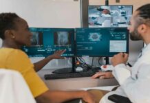
Integrating AI into the radiology workflow can help medical institutions keep pace with an ever-growing stream of medical imaging data. The number of images acquired during common studies like MRI and CT scans has swelled in recent years from tens of images each to hundreds or thousands. It’s a challenge compounded by a rise in the number of patients being imaged.
“It makes for an absolutely overwhelming volume of information to digest,” said Christopher Hess, chair of the UCSF Department of Radiology and Biomedical Imaging. “We’re hoping to use AI to help radiologists better navigate and interact with data, to derive more meaning out of images, and to improve the value of medical imaging for the individual patient.”
Hess says the university also plans to use AI for quantitative imaging, predictive analytics and resource scheduling — giving medical professionals access to insights that were once too time-consuming to calculate or impossible to find without deep learning methods.
UCSF Adopts NVIDIA Clara and DGX Systems DGX-2 at UCSF AI for radiology center
A leading healthcare institution with more than a century of work in radiology, UCSF has long been an innovator in medical imaging. Its radiology department collaborated with industry partners in the 1970s to develop the first MRI systems,now used worldwide to diagnose a variety of conditions, including spinal fractures and brain and heart diseases.
Close to half a million imaging studies are performed at UCSF annually. The medical center has amassed at least a petabyte of imaging data over the years — ranging from small X-ray images to much larger PET/MRI studies. These bigger files can take up gigabytes or now even terabytes of data storage.
Training deep learning models on these massive datasets requires immense computational power. By adopting the high-performance NVIDIA DGX-2, Hess estimates UCSF researchers could cut the time to train AI models from months or days down to hours or even minutes.
The DGX-2 will also enable UCSF to harness multimodal data sources to develop more sophisticated deep learning models to accelerate the radiology workflow.
“We’re interested in integrating data from not only imaging, but also from medical records, genetics and other information sources in the healthcare system,” said Hess. “When we talk about computation at scale, we need access to a high-throughput, highly efficient and computationally sophisticated platform like DGX-2 to accelerate our development cycle.”
UCSF has also adopted the NVIDIA Clara developer toolkit for medical imaging. Its researchers are using the Clara Train SDK to train deep learning models that reconstruct and analyze CT and MRI scans, and the Clara Deploy SDK to optimize integration with the center’s clinical infrastructure.
“We’re really focusing on developing ways in which to implement algorithms from the modality to the reading room,” said Hess. “NVIDIA Clara will be an essential platform to create this ecosystem to implement, validate and use AI algorithms.”
Weaving AI Into the Clinical Workflow
NVIDIA and UCSF are working together to develop AI models that can be deployed into the medical center’s imaging workflow, starting with deep learning models to analyze scans of the brain and liver.
When doctors treat brain cancer patients, MRI scans provide critical information about how a tumor is responding to radiation treatment and chemotherapy. Today, radiologists analyze scans visually with manual tools. AI can instead provide a quantitative measurement, calculating the precise volume of a tumor. By tracking how a tumor’s volume changes from scan to scan, clinicians can better assess how a patient is responding to treatment over time.
The team is also developing an AI model that can segment and measure the left and right lobes of an organ donor’s liver from CT images. These metrics are critical for doctors planning liver transplants from a living donor to a patient, and take up to two hours to delineate and compute by hand. With deep learning, Hess estimates, it could be done in seconds.
UCSF and NVIDIA will also collaborate on tools that could improve the quality, efficiency and reproducibility of medical imaging exams. AI can be used to denoise medical images so that scans can be taken faster and are less susceptible to patient motion during scanning.
Beyond the day-to-day medical imaging workflow, the collaboration will explore predictive analytics tools to provide radiologists and other physicians insights from imaging scans, medical records and even patient sensors.
Additional deep learning algorithms will be created to improve operational efficiency at UCSF, helping its technologists optimize how the medical center’s fleet of imaging scanners is used.


