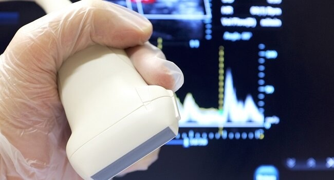
Ultrasound technology has become an invaluable tool in modern medicine. Its imaging capabilities are crucial in aiding diagnosis, guiding interventions, and monitoring patient conditions across specialties.
However, while this diagnostic procedure offers numerous benefits, it also carries potential risks if not used correctly. Adherence to established safety protocols is essential for its safe integration into clinical practice.
This guide aims to cover the key aspects of safe and responsible ultrasound use, providing a useful overview for medical professionals.
Operator Training
Proper training is indispensable for safely harnessing ultrasound’s capabilities. Physicians, sonographers, and other users must complete accredited programs, simulations, and directly supervised practice. Programs, such as Zedu ultrasound training, provide comprehensive knowledge and practical skills covering:
- Physics and technology basics
- Imaging techniques and protocols
- Image optimization and interpretation
- Safe machine operation and transducer handling
- Recognition of imaging artifacts
- Record keeping and documentation
Thorough initial training combined with continuing education enables users to scan patients safely and accurately.
Controlled Scanning Exposure
Ultrasound technology utilizes high-frequency sound waves for imaging. While it doesn’t involve ionizing radiation, cautious exposure management is still essential. Following the ‘as low as reasonably achievable’ (ALARA) principle helps in moderating ultrasound wave exposure based on diagnostic needs. Implementing these methods is crucial:
- Minimizing Power Levels And Duration: Adjust power settings and scanning duration to the lowest sufficient levels for quality imaging to minimize unnecessary exposure.
- Choosing Intermittent Over Continuous Scanning: Opt for intermittent scanning, rather than continuous mode, to reduce overall exposure time.
- Selecting Appropriate Transducer And Frequency: Use the smallest transducer area and highest frequency suitable for the body region and depth required to limit exposure.
These practices ensure that scanning is conducted for the shortest possible time and at the lowest power output necessary, thereby reducing potential bioeffects.
Image Optimization
Proper imaging technique is imperative for examinations to capture necessary views and information with clarity. Optimizing images facilitates accurate interpretation while avoiding the need for excessive scanning or high outputs to improve inadequate images. Adjustment methods include:
- Adjusting Patient Positions: Vary patient positions to achieve the clearest possible view of anatomical structures.
- Enhancing Sound Wave Transmission: Use an adequate amount of gel or saline coupling to maximize sound wave transmission.
- Fine-Tuning Imaging Parameters: Adjust depth, gain, and other ultrasound parameters appropriately for optimal image quality.
- Employing Advanced Imaging Techniques: Utilize spatial compounding, harmonics, and speckle reduction imaging when available to enhance image quality.
- Minimizing Motion Artifacts: Implement patient breath holds or cardiac gating as necessary to reduce motion artifacts in imaging.
By taking these steps, healthcare professionals can obtain excellent diagnostic images, guiding accurate diagnosis and treatment while minimizing the required exposure.
Transducer Safety
Transducers direct ultrasound waves into the body, making proper handling critical. To prioritize safety, the following steps should be observed:
- Regular Inspection: Check for cracks, leakage, overheating, or other damage before each scan.
- Sterility In Invasive Procedures: Utilize sterile sheaths for procedures that are invasive.
- Rigorous Disinfection: Thoroughly disinfect the transducer between patients, following manufacturer protocols.
- Avoiding Sensitive Areas: Be cautious to avoid contact with eyes or lenses during scans.
- Proper Storage: Store transducers hanging upright rather than sitting flat.
Adhering to these safety protocols is critical to protect patients from potential injury and prevent cross-contamination.
Dosimetry Monitoring
Effective tracking of ultrasound exposure is vital for its safe and controlled use. Features like thermal and mechanical indices, on-screen timer displays, and probe surface temperature readouts offer real-time feedback. To further enhance dosimetry monitoring, carry out the following:
- Recording Cumulative Exposure: Log cumulative exposure times, especially for organs more susceptible to bioeffects, such as eyes, heart, and brain.
- Employing Simulation Systems: Use ultrasound simulation systems to monitor scanning techniques and image optimization.
- Analyzing Archived Images: Review archived images to assess exposure habits and the effectiveness of imaging protocols.
Regular monitoring and data analysis help identify opportunities to implement the ALARA principle, allowing adjustments in device settings, techniques, and training to enhance safety.
Safety Testing
Regular safety testing of all ultrasound equipment is necessary to guarantee its proper functioning. This involves comprehensive inspections across the following aspects:
- Electrical Safety: Conduct checks for electrical current leakage, proper grounding, and equipotential bonding to ensure electrical integrity.
- Mechanical Safety: Examine the integrity of transducer housing and the functionality of emergency stops to prevent mechanical failures.
- Acoustic Output Assessment: Verify that power levels are in alignment with manufacturer specifications and intended uses for safe acoustic output.
- Image Quality And Resolution Evaluation: Confirm that resolution, distance calibration, and dead zone are up to par to maintain high standards of image quality.
Conducting these tests regularly contributes to safeguarding both patient and operator safety.
Policies And Documentation
Establishing written policies is fundamental in setting and maintaining appropriate use and safety standards for ultrasound equipment. These policies serve as a guideline for all operators, promoting consistency in practice. Policies should cover:
- Training requirements for operators
- ALARA exposure limits
- Dosimetry monitoring procedures
- Transducer handling and disinfection protocols
- Mandatory safety testing and maintenance
- Appropriate imaging settings for exam types
Clear documentation, including imaging parameters, exposure times, diagnostic justifications, and patient consent, is vital for demonstrating compliance with safety standards and training protocols. This demonstrates adherence to policies and fosters quality assurance in ultrasound practice.
Final Thoughts
Effective ultrasound use in clinical practice relies on comprehensive training, strict adherence to safety protocols, and consistent equipment testing. This approach ensures that this diagnostic tool remains reliable and effective in promoting patient care.

Staying updated with these practices allows healthcare professionals to maximize the benefits of ultrasound, safeguarding patient well-being while enhancing the overall quality of medical care. This commitment to responsible use and continuous improvement supports its role as an asset in modern healthcare.


