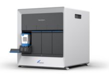
At Mumbai, India’s ACTREC (The Advanced Centre for Treatment, Research & Education in Cancer), neurosurgeons have employed Elekta SonoWand Invite™ in 15 patients with brain tumors since the center acquired the system in July. The integration of sophisticated intra-operative 3D ultrasound and navigation technology in SonoWand provides both the optimal path to the tumor and – with 3D ultrasound – ongoing images of the operative site, enabling the surgeon to safely remove all visible traces of the tumor.
“The goal in any tumor resection surgery is to extract the tumor – and to extract it completely – without causing any harm to healthy brain tissue, which is quite a challenge,” says Aliasgar V Moiyadi, MCh., Assoc. Professor and Consultant Neurosurgeon at ACTREC, part of Mumbai’s Tata Memorial Centre. “SonoWand addresses this challenge by helping you see what you’re removing, how much you’ve removed and how much more you could take out over and above what you could have seen with just your eyes or with the intra-operative microscope.”
In a compact, single-rack solution, SonoWand Invite combines a high-performance ultrasound scanner with a built-in navigational computer and optical tracking system. The system supports several trackable instruments and accessories, such as navigators and ultrasound probes. Planning and navigation displays enable axial, coronal and sagittal views, in addition to a unique user-friendly and intuitive “anyplane view,” which displays the image information in the plane of the actual surgical trajectory.
Although MRI is regarded as the premier intra-operative imaging modality for its image quality, several limitations make intra-operative MRI for brain tumors impractical, Dr. Moiyadi observes.
“An MRI system is very costly, it’s large and cumbersome to work with and it takes too long to do repeated scans during surgery,” he says. “Plus, MRI is not actually ‘real-time,’ – because you see an image, then take the next surgical step, you’re not imaging and resecting simultaneously. SonoWand is an economical and effective solution.”
SonoWand aids “search and destroy” mission against tumors
The SonoWand-assisted tumor resection starts with a diagnostic, multi-plane MRI, during which the patient’s brain and fixed surface fiducial marks are scanned. The MRI images are imported into SonoWand and clinicians register the fiducial marks on both the images and – with an optically-tracked probe – the fiducial marks on the patient’s scalp. Once this registration is complete, every point in the patient’s cranium will have an “address” (x,y,z) in 3D space, which corresponds to the same point in the image.
“When you touch the patient’s head with the probe, you know that this is exactly the point on the image you’re touching,” he says. “And you can, by extrapolation, know that if you go, for instance, five centimeters deeper in a given plane and trajectory, you will reach a particular structure in the brain without actually seeing it, because you can follow it on the image. That is how navigation helps you plan your entry point into the brain.”
The entry point indicates where the surgeon places the craniotomy. By using the navigational capability of the system, the surgeon is able to customize the craniotomy to just the right size. This avoids unnecessarily large or misplaced craniotomies. Once the craniotomy is performed and the dura mater is opened, however, cerebral spinal fluid in which the brain is suspended is released, causing the brain to shift.
“Therefore, the images that were applied preoperatively are now going to be useless because the anatomy has changed,” Dr. Moiyadi explains. “At that point, we turn to the ultrasound capabilities of SonoWand, which provides a specialized probe that takes a sector view of multiple 2D images and fuses them to afford a 3D volume. That 3D volume now serves as your new image with which to navigate. The best part is that the ultrasound probe is already navigating with the SonoWand computer via optical tracking and navigational spheres so we know exactly which part of the brain is being imaged by the probe.”
Following the predetermined path to the tumor, the surgeon can begin to remove or “debulk” the lesion. As debulking progresses, the brain can shift again, necessitating image updates.
“Ultrasound allows us to update that information repeatedly – as many times as we want – without taking too much time,” he says. “In addition, it’s important to note that the SonoWand dedicated cranial ultrasound provides image quality that is far superior to any conventional ultrasound system.”
Exceptional preoperative navigational capabilities and high-quality, navigated ultrasound have clearly benefited patients at ACTREC, helping ensure safer access to brain tumors and more complete removal via real-time imaging, Dr. Moiyadi observes.
“Intra-operative ultrasound – especially one like SonoWand, which is 3D, real-time and combined with navigation – caters to all our needs, including our financial requirements.


